
Employing a novel strategy for growing tissue, MIT and Harvard Medical School scientists have devised a way to label unique molecules of messenger RNA in just a tissue sample and then sequence the RNA.
This strategy delivers a exclusive snapshot of which genes are becoming expressed in distinctive areas of a mobile, and could allow experts to learn significantly more about how gene expression is affected by a cell’s location or its interactions with nearby cells. The technique could also be helpful for mapping cells in the mind or other tissues and classifying them in accordance to their operate.
“Gene expression is 1 of the most fundamental processes in all of biology, and it plays roles in all organic processes, each balanced and illness-relevant. Nonetheless, you will need to know extra than just no matter if a gene is on or off,” claims Ed Boyden, the Y. Eva Tan Professor in Neurotechnology and a professor of organic engineering, media arts and sciences, and brain and cognitive sciences at MIT. “You want to know wherever the gene solutions are situated. You care what mobile styles they are in, which individual cells they participate in roles in, and even which sections of cells they do the job in.”
In a study showing up nowadays in Science, the researchers showed that they could use this procedure to identify and then sequence 1000’s of different messenger RNA molecules in just the mouse brain and in human tumor samples.
The senior authors of the review are Boyden, an investigator at the MIT McGovern Institute and the Howard Hughes Health care Institute George Church, a professor of genetics at Harvard Professional medical University and Adam Marblestone, a previous MIT study scientist. The paper’s direct authors are Shahar Alon, a former MIT postdoc who is now a senior lecturer at Bar-Ilan College Daniel Goodwin, an MIT graduate university student Anubhav Sinha ’14 MNG ’15, an MIT graduate pupil Asmamaw Wassie ’12, PhD ’19 and Fei Chen PhD ’17, who is an assistant professor of stem mobile and regenerative biology at Harvard College and a member of the Broad Institute of MIT and Harvard.
Tissue enlargement
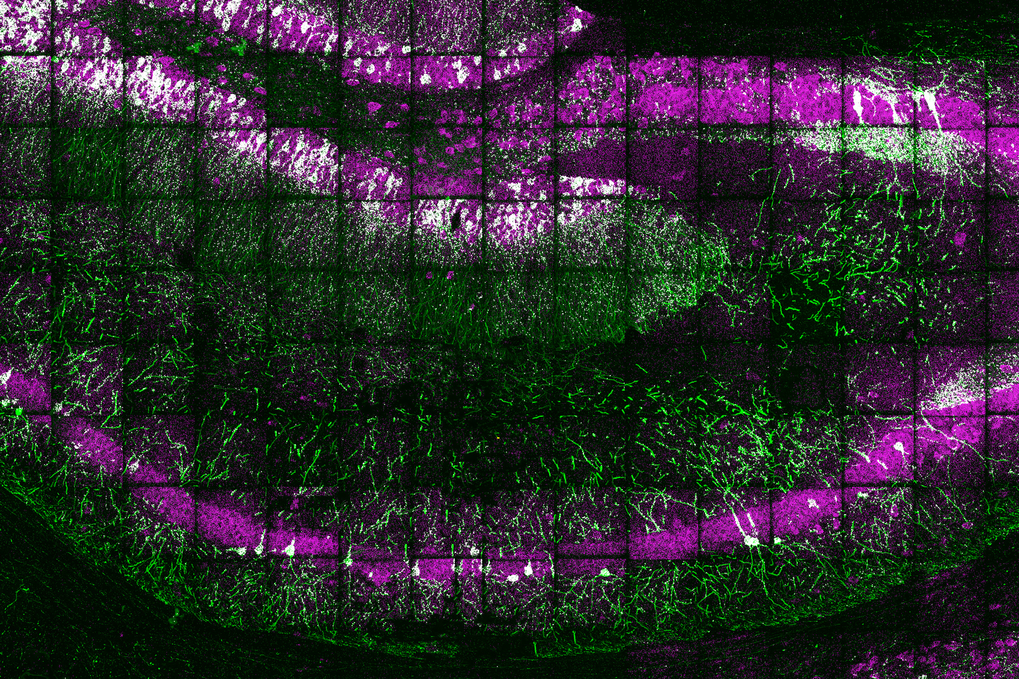
The new sequencing procedure builds on a method that Boyden’s group devised in 2015 for expanding tissue samples and then imaging them. By embedding water-absorbent polymers into a tissue sample, scientists can swell the tissue sample whilst keeping its overall organization intact. Working with this method, tissues can be expanded by a component of 100 or far more, allowing for scientists to obtain extremely significant-resolution images of the mind or other tissues applying a standard gentle microscope.
In 2014, Church’s lab made an RNA sequencing procedure acknowledged as FISSEQ (fluorescent in situ sequencing), which will allow countless numbers of mRNA molecules to be positioned and sequenced within just cells developed in a lab dish. The Boyden and Church labs made the decision to be a part of forces to mix tissue enlargement and in situ RNA sequencing, building a new approach they simply call expansion sequencing (ExSeq).
Expanding the tissue just before accomplishing RNA sequencing has two most important positive aspects: It offers a better-resolution seem at the RNA in cells, and it helps make it simpler to sequence individuals RNA molecules. “When you individual these molecules in the expanding sample, and go them away from every other, that gives you additional home to basically accomplish the chemical reactions of in situ sequencing,” Marblestone suggests.
At the time the tissue is expanded, the scientists can label and sequence 1000’s of RNA molecules in a sample, at a resolution that allows them to pinpoint the molecules’ destinations not only within cells but inside of distinct compartments these kinds of as dendrites — the small extensions of neurons that acquire communications from other neurons.
“We know that the place of RNA in these little locations is vital for mastering and memory, but until eventually now, we failed to have any way to evaluate these areas due to the fact they are very tiny, on the purchase of nanometers,” Alon states.
Utilizing an “untargeted” edition of this system, this means that they are not wanting for precise RNA sequences, the researchers can turn up countless numbers of distinct sequences. They estimate that in a provided sample, they can sequence among 20 and 50 percent of all of the genes current.
In the mouse hippocampus, this technique yielded some stunning benefits. For a single, the scientists found mRNA containing introns, which are sections of RNA that are usually edited out of mRNA in the nucleus, in dendrites. They also found out mRNA molecules encoding transcription aspects in the dendrites, which may possibly assistance with novel forms of dendrite-to-nucleus conversation.
“These are just illustrations of factors that we by no means would have gone on the lookout for deliberately, but now that we can sequence RNA exactly in which it is in the neuron, we’re ready to investigate a whole lot extra biology,” Goodwin says.
Mobile interactions
The scientists also confirmed that they could investigate gene expression in a additional focused way, hunting for a unique established of RNA sequences that correspond to genes of interest. In the visual cortex of the mouse, the researchers used this tactic to classify neurons into unique varieties primarily based on an examination of 42 distinct genes that they specific.
This technological know-how could also be valuable to assess a lot of other forms of tissues, such as tumor biopsies. In this paper, the scientists analyzed breast cancer metastases, which comprise several distinct cell sorts, including most cancers cells and immune cells. The research unveiled that these mobile forms can behave in different ways relying on their location inside a tumor. For instance, the researchers found that B cells that were around tumor cells expressed selected inflammatory genes at a larger stage than B cells that ended up farther from tumor cells.
“The tumor microenvironment has been examined in a lot of distinctive contexts for a extensive time, but it’s been hard to study it with any depth,” Sinha suggests. “A most cancers biologist can give you a checklist of 20 or 30 marker genes that will discover most of the mobile styles in the tissue. Here, due to the fact we interrogated 297 various RNA transcripts in the sample, we can request and remedy much more in-depth queries about gene expression.”
The scientists now program to more analyze the interactions between cancer cells and immune cells, as perfectly as gene expression in the brain in healthful and illness states. They also strategy to extend their tactics to enable them to map supplemental styles of biomolecules, these kinds of as proteins, alongside RNA.
The investigate was funded, in section, by the Nationwide Institutes of Wellbeing and the Countrywide Science Foundation, as very well as by Lisa Yang, John Doerr, the Open Philanthropy Venture, Cancer Study Uk, the Chan Zuckerberg Initiative Human Cell Atlas pilot software, and HHMI.